 English





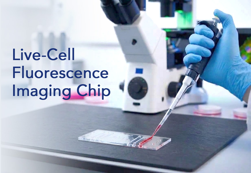
Live-Cell Fluorescence Imaging Chip
CIC-6F
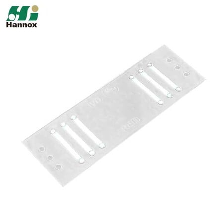
Six-channel microfluidic imaging chip can use with a 170 µm optical bottom for confocal fluorescence microscopy, live-cell observation, blood cells, and micro-volume samples operation.
Fluorescence Microscopy Chip
Bring Small Samples into an Imaging-Ready Microchannel
Live-Cell Fluorescence Imaging Chip is a six-channel microfluidic observation chip developed for fluorescence and high-magnification microscopy. Samples remain inside defined microchannels, allowing researchers to work with small volumes while keeping cells or blood cells in an imaging-friendly format.
Optical Bottom Designed for Confocal Microscopy
The chip uses a 170 µm optical bottom, corresponding to the standard 0.17 mm coverslip thickness commonly used for high-resolution microscopy. This design supports confocal fluorescence microscopy and other high-magnification imaging workflows.
Six Independent Channels for Efficient Experimental Design
Each chip contains six short channels, allowing up to six independent experiments or conditions on a single slide-format device. Standard pipettes can be used to introduce cell suspensions or blood samples, making the chip easy to integrate into routine laboratory workflows.
FEATURE
- Compatible with confocal fluorescence microscopy and high-magnification observation.
- 170 µm optical bottom designed around standard microscopy optics.
- Six independent microfluidic channels on one chip.
- Supports observation, image capture, and video recording of rare or micro-volume samples.
- Suitable for high-sensitivity immunofluorescence staining workflows.
- Supports live-cell and blood-cell observation and research.Use small sample volumes to help conserve cells and reagents.
- Use small sample volumes to help conserve cells and reagents.
- Samples are contained within microchannels rather than spread across a conventional flat slide.
- Compatible with standard pipettes for introducing cell suspensions or blood samples.
- Can be used for cell and microbial experiments within the microchannels.

APPLICATION
Confocal Fluorescence Microscopy
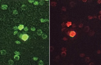
Designed for high-resolution fluorescence imaging where optical compatibility and controlled sample placement are important.
Immunofluorescence Studies
Suitable for high-sensitivity immunofluorescence staining with limited sample volumes.
Live-Cell and Blood-Cell Observation
Supports direct microscopic observation of live cells and blood cells, including image capture and video recording.

Micro-Volume and Precious Samples
The microchannel format helps reduce sample and reagent consumption, making it useful when cells or reagents are limited.
Microbial and Cell-Based Experiments
The channels can be used as a compact environment for cell or microorganism observation and other microscopy-centered experimental workflows.
SPECIFICATION
| Item | Specification |
|---|---|
| Outer dimensions | 75 × 25 × 0.85 mm |
| Number of channels | 6 |
| Channel dimensions | 2 × 15 mm |
| Optical bottom thickness | 170 µm (0.17 mm) |
| Inlet / outlet hole size | Ø 2 mm each |
| Material | Polymer / Glass |
| Color | Transparent |
| Microscopy compatibility | Confocal fluorescence microscopy and high-magnification microscopy |
| Sample loading | Compatible with standard pipettes |
| Package | 12 pcs / box |
DIRECTION FOR USE
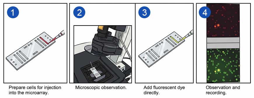
A general imaging workflow is provided below. Sample preparation, staining, incubation, and imaging conditions should be selected according to the experimental protocol.
- Prepare the cell suspension, blood sample, microorganism sample, or stained specimen according to the study protocol.
- Using a standard pipette, introduce the sample through the inlet and allow the microchannel to fill without trapping bubbles.
- Confirm that the sample is positioned in the observation region of the selected channel.
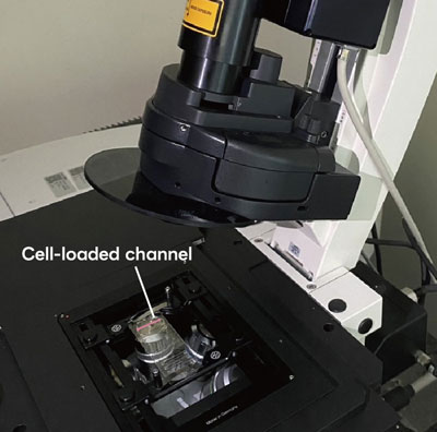
- Place Live-Cell Fluorescence Imaging Chip on the microscope stage and selecting the appropriate objective and imaging mode.
- For fluorescence experiments, use excitation, emission, exposure, and acquisition settings established for the fluorophore and microscope system.6. Capture images or video as required by the experiment.
- Capture images or video as required by the experiment.
- Use the remaining channels for additional replicates, conditions, or samples as appropriate.
- After completion, handle and dispose of the used chip according to the laboratory's biological-safety procedure.
Note: Recommended loading volume and incubation conditions are protocol-dependent.
FAQ
| 1. Is Live-Cell Fluorescence Imaging Chip compatible with confocal fluorescence microscopy? Yes. Live-Cell Fluorescence Imaging Chip is specifically designed to support confocal fluorescence microscopy and high-magnification observation. |
| 2. What is the optical bottom thickness? The chip uses a 170 µm, or 0.17 mm, optical bottom designed around standard high-resolution microscopy optics. |
| 3. How many experiments can be run on one chip? Live-Cell Fluorescence Imaging Chip contains six independent microfluidic channels, allowing up to six separate experimental conditions or replicates on one chip. |
| 4. Can I load samples with a standard laboratory pipette? Yes. The inlet and outlet design allows cell suspensions or blood samples to be introduced using a standard pipette. |
| 5. What types of samples can be observed? The product is intended for cell and blood-cell observation and can also be used for microbial experiments in microchannels. It is particularly useful for rare or micro-volume samples. |
| 6. Is Live-Cell Fluorescence Imaging Chip suitable for immunofluorescence? Yes. The product is described as suitable for high-sensitivity immunofluorescence staining and fluorescence imaging. |
| 7. How does the microchannel format help when samples are limited? Because the observation takes place in a defined microchannel using a small sample volume, researchers can reduce the amount of cells and reagents required compared with workflows that use larger sample areas or volumes. |
| 7. Can Live-Cell Fluorescence Imaging Chip be used for video or dynamic observation? Yes. The product supports image capture and video recording. Time-dependent or live-cell imaging can be performed when the microscope, environmental conditions, and experimental protocol are appropriate. |
Welcome Private Label, OEM/ODM, Distribution, and Agency Partnerships
For any further question ( Minimum Order Quantity, Prices or Domestic Distributor), please fill out the inquiry form, we will get back to you as soon as possible. Let us work together to make your product come true and hit the market successfully.